


 33 / 70

55°C for about 12 hours, and
then cycled between roughly
5° and 20°C during the
EOIM-3 exposure period. Sputtered films of pure MoS
2
[5]
representing the historical, columnar growth morphol-
ogy typical of undoped films were evaluated alongside
structurally modified films that were co-sputtered with
antimony oxide (MoS
2
/Sb
2
O
3
)
[6]
, or deposited in multi-
layers with nickel (MoS
2
/Ni)
[7]
. In addition to LEO expo-
sure, films were also exposed in a terrestrial facility
[8]
using AO energy per atom and UV exposures designed
to emulate the LEO environment. In this case, films expe-
rienced an AO fluence of 1.97×10
20
atoms/cm
2
(about 15%
less than in the space flight) during an exposure of 25.28
hours. Several identical sets of samples were produced for
EOIM-3 so that samples could be examined after LEO ex-
posure, after AO exposure in the laboratory, and after des-
iccated storage on earth for the
same time period.
More recently, a compos-
ite MoS
2
film was exposed to
LEO during the Materials on
the International Space Sta-
tion-7 (MISSE-7) experiment.
Samples were installed on the
ISS on November 23, 2009,
during STS-129 (another At-
lantis mission), and recovered
on March 1, 2011, during
STS-133 (shuttle Discovery),
accumulating 463 days of
exposure. MISSE-7 samples
were exposed to 3.60×10
21
atoms/cm
2
, roughly 14.4 times the total fluence of the
earlier EOIM-3 experiment. Composite MoS
2
films were
co-sputtered with antimony oxide and gold
(MoS
2
/Sb
2
O
3
/Au)
[9]
, and were enclosed in “suitcase style”
passive experiment containers (PECs), shown in Fig. 1, as
part of Sandia’s Passive ISS Research Experiment (SPIRE)
on MISSE-7. These LEO exposure experiments were con-
sidered passive, while active experiments were led by B.
Krick and W.G. Sawyer at the University of Florida, where
data from sliding wear tests was collected on orbit and
downloaded directly from space.
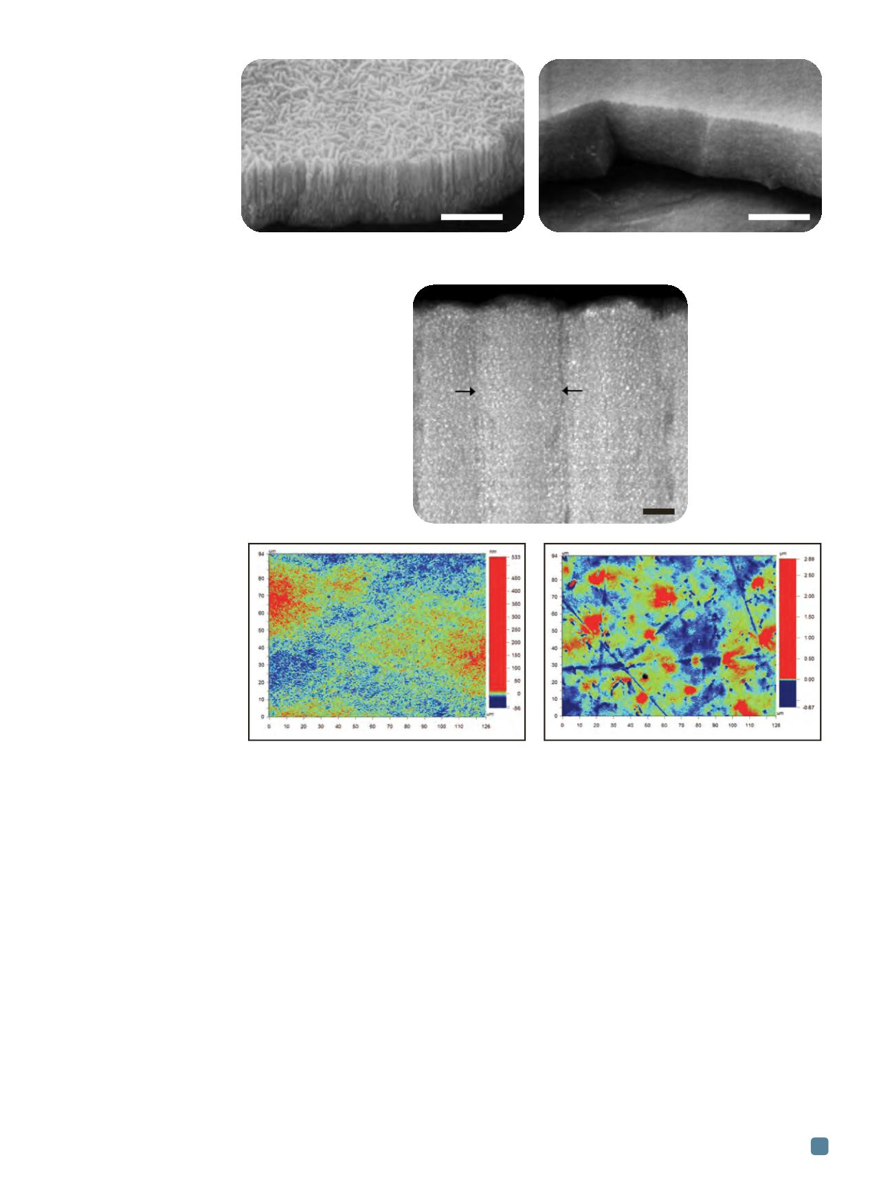
Figure 2 shows the morphology of MoS
2
films at frac-
tured edges created intentionally by plastically deforming
the substrate through indentation with a hardened steel
ball. The columnar morphology of the pure MoS
2
film is
contrasted with the smooth fracture morphology of the
MoS
2
/Sb
2
O
3
film. Doping of MoS
2
films interrupts the
columnar growth of pure MoS
2
, and tends to create films
with more equiaxed grains, and hence reduced porosity.
Figure 3 shows a cross-sectional HAADF-STEM image
of the MoS
2
/Sb
2
O
3
/Au film. This film is a composite with
nanoscale Au particles distributed throughout the amor-
phous MoS
2
/Sb
2
O
3
matrix. Surface roughness changes ac-
companying exposure of MoS
2
/Sb
2
O
3
/Au films to LEO
during the MISSE-7 mission are illustrated in Fig. 4. RMS
roughness increased significantly from as-deposited films
changing from 8.0±0.3 to 34.0±7.9 nm after LEO exposure.
This increase in roughness is likely due to the effects of
heterogeneous erosion of the film by AO, as well as poten-
tial impacts of micrometeoroids or other space debris.
Typical friction coefficient behavior for MoS
2
films is
shown in Fig. 5(a). Friction traces for the MoS
2
/Sb
2
O
3
/Au
films show that sliding begins with a friction coefficient
that is elevated with respect to the steady-state value es-
tablished after several cycles of sliding contact. This initial
friction coefficient is attributed to various chemical and
ADVANCED MATERIALS & PROCESSES •
MAY 2014
33
Fig. 2 —
Fracture surface morphology of pure MoS
2
exhibits a columnar growth morphology (a), and dense
MoS
2
/Sb
2
O
3
films (b) exposed to LEO on EOIM-3.
(a) (b)
Fig. 3
—
Cross-sectional
HAADF-STEM image
of the MoS
2
/Sb
2
O
3
/Au
film. Brighter Au
nanoparticles are
distributed throughout
the film. Arrows show
intercolumnar
boundary.
Fig. 4 —
Contour maps from optical interference profilometry of the MoS
2
/Sb
2
O
3
/Au film in as-deposited
condition (left) with RMS roughness of 8.0±0.3 nm and after exposure to LEO during MISSE-7 (right) with
RMS roughness of 34.0±7.9 nm.
1 µm
1 µm
25 nm