


 12 / 50

A D V A N C E D M A T E R I A L S & P R O C E S S E S | A P R I L 2 0 1 7
1 2
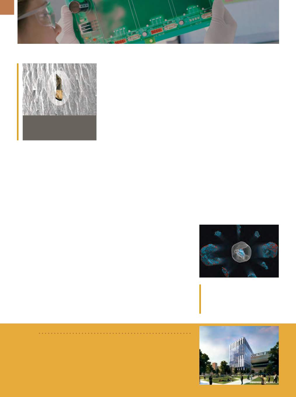
TOOTH-INSPIRED MATERIAL
TAKES THE SHAKE
Researchers at the University of
Michigan, Ann Arbor, designed a rig-
id material with exceptional vibration
absorption capabilities modeled after
tooth enamel. Enamel can withstand
decades of impact due to its structure:
columns of ceramic crystals infiltrat-
ed with a matrix of proteins set into a
protective coating. When the stiff na-
noscale columns bend under stress
and press against the soft polymer
surrounding them, friction occurs, and
the large contact area dissipates ener-
gy that would otherwise be damaging.
To recreate this structure, researchers
grew zinc oxide nanowires on a chip
then poured on layers of polymer. With
each layer, the chip was spun to spread
the polymer, then baked. It took 40
layers to build up a single microme-
ter of synthetic enamel, and to further
increase the material’s thickness, the
whole process was repeated up to 20
times. The painstaking layering ensures
that surfaces are perfectly mated—even
nanoscale gaps between the polymer
and ceramic reduce friction intensity.
Computer modeling confirms that
vibrational forces are diffused through
the polymer and column interaction,
and the synthetic enamel prevents
damage almost as well as real tooth
enamel. “Artificial enamel is better than
solid commercial and experimental ma-
terials that are aimed at the same vibra-
tion damping,” explains Nicholas Kotov,
professor of chemical engineering. “It’s
lighter, more effective, and perhaps less
expensive.” Kotov believes the material
could be deployed in airplanes and oth-
er environments in which vibrations are
inescapable.
umich.edu.3D ATOM MAP REVEALS
BOUNDARY ISSUES
Using advanced electron micros-
copy and powerful reconstruction al-
gorithms, physicists at the University
of California, Los Angeles mapped the
coordinates of more than 23,000 in-
dividual atoms in an iron-platinum
nanoparticle. The team identified and
located more than 6500 iron and 16,600
platinum atoms in the particle and de-
termined that the atoms are arranged
in nine grains, each of which contains
different ratios of iron and platinum at-
oms. They also showed that atoms clos-
er to the interior of the grains are more
regularly arranged than those near the
surfaces and that the grain boundaries
Identification of the precise 3D coordi-
nates of iron, shown in red, and platinum
atoms in an iron-platinum nanoparticle.
Courtesy of Colin Ophus and Florian
Nickel.
BRIEF
The Sir Henry Royce Institute for Advanced Materials,
UK, received a government
grant of £128 million (approximately $156 million) to support precision equipment
and construction of state-of-the-art facilities, including a new hub at
The Univer-
sity of Manchester.
The institute will explore nine key areas grouped into four
themes—energy, engineering, and functional and soft materials—and focus on the
commercialization of fundamental advanced materials research.
www.royce.ac.uk.
are more disordered. When research-
ers fed the coordinates into quantum
mechanics calculations, they observed
abrupt changes in magnetic properties
at the grain boundaries.
“Understanding the 3D structures
of grain boundaries is a major chal-
lenge in materials science because they
strongly influence the properties of ma-
terials,” explains Jianwei “John” Miao,
professor of physics and astronomy.
“Now we are able to address this chal-
lenge by precisely mapping out the 3D
atomic positions at the grain boundar-
ies for the first time.” The work makes
significant advances in characterization
capabilities and the understanding of
structure-property relationships, and
could find broad application in materi-
als science as well as physics, chemis-
try, and nanotechnology.
ucla.edu.EMERGING TECHNOLOGY
Illustration of the Henry Royce Institute. Courtesy of NBBJ and Vyonyx.
T. rex
tooth and an electronmicro-
scope image of the tooth enamel.
Courtesy of Kotov Lab, University of
Michigan.