


 10 / 50

2014 is International Year of Crystallography
One hundred years ago,
x-rays were used for the first time
to determine the crystal structure
of materials. The pioneering work
of Max von Laue, for which he
won the Nobel Prize in physics,
was followed by a joint award in
1915 (also in physics) to father and
son Sir William Henry Bragg and
William Lawrence Bragg. Since
then, more than 25 Nobel Prizes
were awarded in areas related to x-
ray diffraction, such as crystallog-
raphy, electron microscopy, and
topics related to the structure of
matter and its properties.
The United Nations pro-
claimed 2014 to be the Interna-
tional Year of Crystallography as
a way to commemorate not only
the achievements of von Laue and
the Braggs, but also to mark the 400th anniversary of Johannes Kepler’s observation of
symmetry in ice crystals, which sparked wider study of the role of symmetry in matter.
The discovery of x-rays in the late 19th century transformed the field of crystallogra-
phy, which previously relied on classifying crystals based on morphological appearance.
The interaction of x-rays with crystals showed that x-rays are electromagnetic waves with
a wavelength of about 10
-10
m, and that the internal structure of crystals was spatially pe-
riodic over long distances. Since then, crystallography has become a basic discipline of
many branches of physical and biological sciences, including materials science and metal-
lurgy. Understanding the role of defects in crystal lattices and how they affect the deforma-
tion of metals has revolutionized the design of materials for the infrastructure of the
industrialized world.
www.iycr2014.org,
www.llnl.gov.WPI leads project to design lightweight metals for military vehicles
Worcester Polytechnic Institute (WPI), Mass., is the lead institution on a $7.4 million,
multi-university award from the U.S. Army to support development of new metallurgical
methods and lightweight alloys to help build more effective and durable military vehicles
and systems. WPI will receive $2.1 million through the two-year award to develop data-
bases and computer modeling techniques that make it possible to predict nanoscale prop-
erties of lightweight alloys (primarily aluminum, titanium, and magnesium), and to use
these computational tools to design and test new alloys for specific military applications.
“The military wants new alloys that are strong enough to be used structurally, tough
enough to function as armor, and light enough to improve the mobility and fuel economy
of vehicles,” says Richard Sisson, director of WPI’s Materials Science and Engineering Pro-
gram and principal investigator for the Army award.
Sisson is working with co-principal investigators Diran Apelian, director of the Metal
Processing Institute (MPI), and Makhlouf Makhlouf, director of the MPI’s Aluminum Cast-
ing Research Center. They are using a variety of modeling techniques, including thermo-
dynamic and kinetic models plus laboratory studies, to predict the microstructure and
microchemistry of new alloys. A variety of metallurgical processes, including heat treating,
are also being studied. From this work, modelling tools that can be used to improve exist-
ing alloys or design new alloys with the desired properties are being derived.
The award also includes a focus on high-strength magnesium alloys used by the mili-
briefs
FEI Co.,
Hillsboro, Ore., installed a
suite of high-end electron
microscopes at
Simon Fraser
University’s (SFU) Centre for Soft
Materials,
Vancouver, B.C.
Systems include the Tecnai Osiris
transmission electron microscope
and Helios DualBeam scanning
electron microscope/focused ion
beam. The systems join several
other FEI microscopes in the 4D
LABS facility where the new
multimillion dollar center is
housed.
www.fei.com,
www.sfu.ca.
Scientists use the Helios
NanoLab DualBeam at Simon
Fraser University.
JEOL USA Inc.,
Peabody, Mass.,
launched an image contest to
showcase some of the best work
of its electron microscope users. A
winning entry is being selected
each month this year, judged by
JEOL’s SEM and TEM applications
teams for both technical and
artistic qualities. All submissions
are part of an online gallery with a
description of the sample and
technique used. The first winner—
Nano Bling—was submitted by
Colin Davis of
Rocky Mountain
Laboratories
in Golden, Colo. The
image, taken with an older model
field emission SEM installed in the
1990s, was chosen for its
contrasting textures and
amorphous threads leading to an
ordered sharply
angular crystal.
www.jeolusa.com.
Nano Bling.
Courtesy of
Rocky Mountain
Laboratories.
ADVANCED MATERIALS & PROCESSES •
APRIL 2014
10
T
ESTING
C
HARACTERIZATION
news
industry
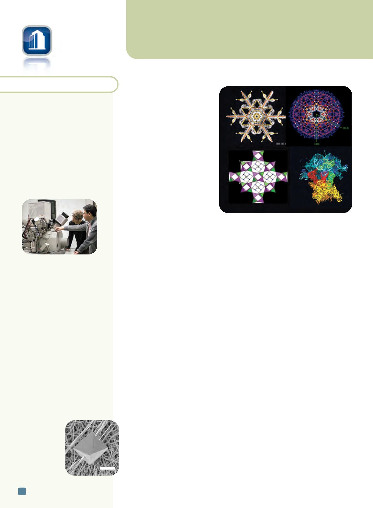
From left, clockwise, crystal structures of a snowflake,
free electron laser, zeolite, and ribosome. Courtesy of the
International Union of Crystallography.
5
m
m