
48
Specimen Name:
Center for Electron Microscopy and Analysis (CEMAS)
Vital Statistics:
• New materials characterization epicenter at The Ohio State
University features $28 million worth of equipment, including
10 FEI electron microscopes.
• The 20,000-sq-ft center includes two electron microscopes
from FEI’s Titan line, optimized to perform atomic scale
analysis. Three additional transmission electron
microscopes, two dual-beam focused ion beam instruments,
and three scanning electron microscopes are also available.
In addition, the center hosts two x-ray diffractometer systems,
facilities for nanoindentation, and an extensive array of sample
preparation facilities.
• Microscopes live in a pristine environment in this purpose-
built facility, free from the effects of vibration, fluctuations
in temperature and humidity, and magnetic fields.
Success Factors:
• Instrumentation and expertise at CEMAS enables analysis of
materials beyond metals and ceramics. Researchers from both
industry and academia can investigate cellular structures of
polymers, tissues, organic membranes, nanoparticles, and gels.
• Due to CEMAS’ direct connection to OARnet’s 100 gigabits-
per-second network, any organization connected to OARnet’s network
can purchase time and directly operate the instruments from a
remote location with no perceptible delay.
• The center aims to revolutionize how scanning electron
microscopy is taught to students. “Since CEMAS’ microscope
collection can be controlled from outside the room, we
no longer need to teach three students at a time,” says
director David McComb. “We can teach 25 or 30 students at
once and we can give them control of the microscope so
everyone can see what’s going on.”
About the Innovators:
An expert in the development and application of
electron energy-loss spectroscopy, David McComb
was recruited to Ohio State to design and lead CEMAS.
Working closely with McComb as academic advisors
are Professors Hamish Fraser and Michael Mills.
Designing the center to help each microscope meet
its exact specification took as long as the physical
construction of converting an old mattress factory into
a world-class research facility. Required modifications include
controlling for temperature to plus or minus 0.1°C without creating air currents,
avoiding vibrations, and removing magnetic fields wherever possible.
What’s Next:
The facility is now open for use by interested parties from academia and industry alike, with
remote microscope operation possible. Book equipment time by contacting CEMAS.
Contact Details:
Center for Electron Microscop
y and Analysis (CEMAS)1305
Kinnear Rd., Colu mbus, OH 43212614/643-3110,
cemas@osu.edu,www.go.osu.edu/electron
Welcome to
Success Analysis,
a
new department featuring in - the- field
reporting on significant advances in
materials science. In each issue, we
will highlight a state - of - the - art
facility, product, research project, or
organization making a noteworthy
impact on the world of materials
engineering.
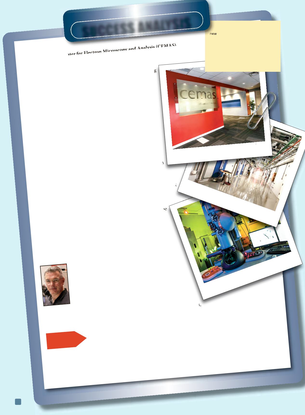
Ohio State's elite electron microscopy
facility celebrated its grand opening on
September 18, 2013.
Specialized systems control for
temperature, humidity, vibration, and
magnetic fields.
CEMAS is home to $28 million worth of
equipment housed in a pristine
environment.
SucceSS AnAlySiS
ADVANCED MATERIALS & PROCESSES •
JANUARY 2014
David McComb


